Breast density is like a double-edged sword for women who have it. Not only does it increase the risk of breast cancer five-fold relative to women with low-density tissue, it hides cancers on mammograms, leading to lower accuracy of mammography. In fact, up to 50% of cancers in women with dense breasts are missed due to lesion obscuration. Because of this, women in this category should be eligible for ultrasound or an MRI--with the related costs covered by health insurance companies. Data available from countries that provide the additional imaging suggest that tumors could be detected in early stages more often, potentially saving thousands of American women’s lives every year.
To address dense breast tissue problems, mammogram equipment manufacturers developed 3-D mammography. The idea is to take multiple X-ray images of the breast at various angles over an arc, and use computer programs to reconstruct multiple “slice” images across the breast. Even this advancement has not completely solved the problem. In some instances, 3-D mammograms have missed breast cancers the size of a tennis ball. These occurrences led to the passage of federal legislation in 2019 that requires radiologists to inform women if they have high breast density so they can consider alternatives and/or additional tests to screen for breast cancer. Unfortunately, insurance coverage for testing beyond mammography is not guaranteed.
Several additional diagnostic options are available to women with dense breast tissue; each with positives as well as downsides:
The Society of Breast Imaging (SBI) and the American College of Radiology (ACR) suggest supplemental ultrasonography. Ultrasound has high sensitivity to detect breast cancer regardless of breast tissue density. However, the procedure is sonographer-dependent and has low specificity, resulting in high false positive rates. A systematic review of the literature showed that the unnecessary biopsy rate was unacceptably high at 92%. In other words, for every 100 women biopsied following a positive ultrasound, only eight would be detected with breast cancer.
Several organizations, including American Cancer Society (ACS), National Comprehensive Cancer Network (NCCN), and SBI, recommend breast magnetic resonance imaging (MRI) for women with a lower than 20% lifetime risk of breast cancer. This imaging technique, which is not impacted by dense breast tissue, uses strong magnetic fields and a contrast agent (gadolinium) injected intravenously. Currently, lower cost “abbreviated” MRIs (a quicker version of the full MRI) are being developed. An ongoing clinical trial is comparing performance of an abbreviated MRI to 3-D mammography in women with dense breast tissue. However, like ultrasounds, MRIs also are not specific and can result in high false positive rates and unnecessary biopsies. Nevertheless, MRI remains the best option for women with high-density breast tissue.
In addition, a number of techniques fall under the nuclear imaging category, which consist of a radioisotope labeled to a cancer-targeting agent that is injected into a patient's blood. Radiation emission from the radioisotope creates images once the targeting agent has been allowed sufficient time to reach the cancer via blood circulation. While these techniques are not impacted by breast density, they are expensive and expose the patient to radiation and, hence, are not suitable for screening average risk patients. However, these techniques are valuable for detecting cancer in high-risk patients and for follow-up patients while they are being treated. Since these techniques involve a cancer-targeting agent, they are more cancer specific and result in fewer false positives compared with ultrasound and MRI.
Each of these options is appropriate in certain situations. Fortunately, diagnostic tools are constantly being refined, and new ones developed. The problem, of course, is a lack of willingness among health insurers to assume the costs of these vital options for women identified as having dense breast tissue, and for lawmakers to insist that they are covered. Mammography alone means possibly missing evidence of breast cancer in half of American women, essentially, 83.5 million wives, mothers, daughters and sisters. There exists enough justification for current legislation to be considered for revision to give them a greater chance for early detection, treatment and recovery.
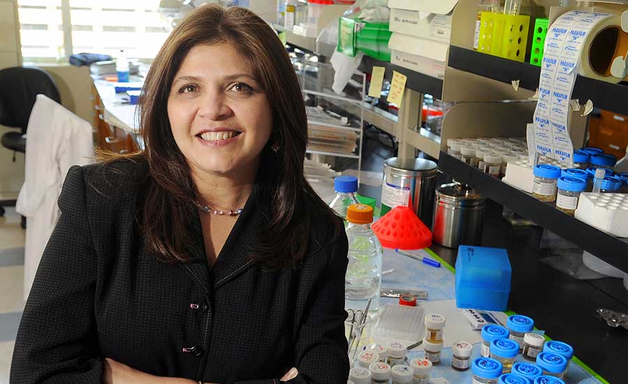
Pinku Mukherjee is the Irwin Belk Endowed Professor of Cancer Research at UNC Charlotte. Research in her laboratory has been and is currently funded through grants from National Cancer Institute, U.S. Department of Defense, Susan G. Komen Foundation and the Pancreatic Cancer Action Network/American Association of Cancer Research.
References
Screening ultrasound as an adjunct to mammography in women with mammographically dense breasts. Am J Obstet Gynecol 2015, 212(1): 9-17
First experiences in screening women at high risk for breast cancer with MR imaging, Breast Cancer Res Treat 2000, 63(1): 53-60
Modern Breast Cancer Detection: A Technological Review, International Journal of Biomedical Imaging, 2009, doi: 10.1155/2009/902326
Nazari, S.S., Mukherjee, P. An overview of mammographic density and its association with breast cancer. Breast Cancer 25, 259–267 (2018). https://doi.org/10.1007/s12282-018-0857-5